Radiological Imaging
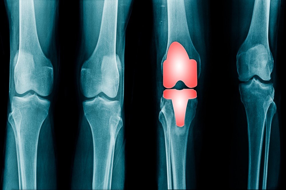
A standard procedure of radiological imaging for knee problem includes the lateral and anterior view of the knee. The lateral imaging allows evaluation of the vertical structure of the patella as in the case of patella alta, which indicates Sinding-Larsen syndrome, lateral patella subluxation, or chondromalacia. The lateral radiograph also indicates the measurement of patellar tilt to rule out the condition of dislocation or patellofemoral joint pain. The axial view reveals the sulcus and congruence angles for the presence of patellar subluxation. Kinematic MR imaging is better than axial radiographs to evaluate the need of patella realignment surgical procedure for the patient. [5]
References
https://www.ncbi.nlm.nih.gov/pmc/articles/PMC3684623/
https://pubmed.ncbi.nlm.nih.gov/23508515/
https://qims.amegroups.com/article/view/13074/13449
https://www.ncbi.nlm.nih.gov/pmc/articles/PMC3553228/
https://pubmed.ncbi.nlm.nih.gov/17669391/