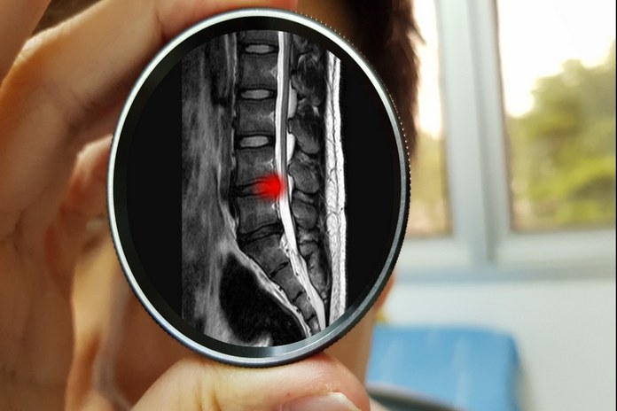
Spinal Stenosis Diagnosis

Your doctor may be able to diagnose spinal stenosis based on your symptoms and a physical exam. He or she may also order tests such as neuronal studies, an X-ray, MRI, or CT scan to help determine the cause of your symptoms. (5)
Physical examination
Spinal stenosis is typically diagnosed through a physical examination, which includes a review of the patient’s medical history and a physical examination of the spine. The neurophysician will look for any signs of narrowing in the spinal canal and will test reflexes and muscle strength in the legs. If spinal stenosis is suspected, the doctor may order additional tests, such as an X-ray or MRI.
Imaging tests
Imaging tests, such as x-rays, CT scan and MRI, are used to diagnose spinal stenosis.
X-rays – X-rays are often used to diagnose spinal stenosis. The images produced by x-rays help doctors see the size and shape of the spinal column. This information can help them determine if there is any narrowing present. X-rays can also help doctors identify other conditions that may be causing the symptoms of spinal stenosis. For example, they may be able to see if a tumor or herniated disk is putting pressure on the spine.
CT scan – Spinal stenosis is often diagnosed with a CT scan. A CT scan uses X-rays to create images of the body. It can help to diagnose spinal stenosis by looking at the bones and tissues around the spine. It can help your neurophysician see if there is any damage to the bones or tissues around the spinal cord.
MRI – A magnetic resonance imaging (MRI) scan is a common diagnostic tool used to identify spinal stenosis. MRI uses a powerful magnetic field and radio waves to create detailed images of the inside of the body. This allows doctors to see if there is any narrowing or damage to the spinal cord or other tissues. If spinal stenosis is suspected, an MRI may be recommended to get a more accurate diagnosis.
Neuronal studies
There are a number of neuronal study tests that can be used to diagnose spinal stenosis including;
Myelogram – One common test is called a myelogram. This test involves injecting dye into the spinal fluid and then taking images of the spine. This can help identify any areas where there is compression on the spinal cord or nerves. This test can also help determine if surgery is needed to treat spinal stenosis.
Needle electromyography – A needle electromyography (EMG) test can help diagnose spinal stenosis. During this procedure, a thin and sterile needle is inserted into the muscles to measure the electrical activity. This can help identify whether there is damage to the nerves and how severe it is.
Nerve conduction study – It is a diagnostic test used to measure the speed of electrical impulses along nerves. This test can help to identify nerve damage, which may be caused by conditions such as spinal stenosis. Nerve conduction study is typically performed on the arm and leg, and involves using electrodes to stimulate the nerve and measure the response. This test can help to determine if there is compression of the nerve due to narrowing of the spinal canal, which can cause pain, numbness, and weakness.
Somatosensory evoked potentials – In some cases, spinal stenosis may not be detectable through standard imaging tests such as an MRI or CT scan. In this case, a physician may order a somatosensory evoked potentials (SSEP) test to help diagnose the condition. SSEP measures the electrical activity in the nerves that travel from the brain to the spinal cord. This test can help determine if there is compression or damage to these nerves.
