Stages of Gynecomastia
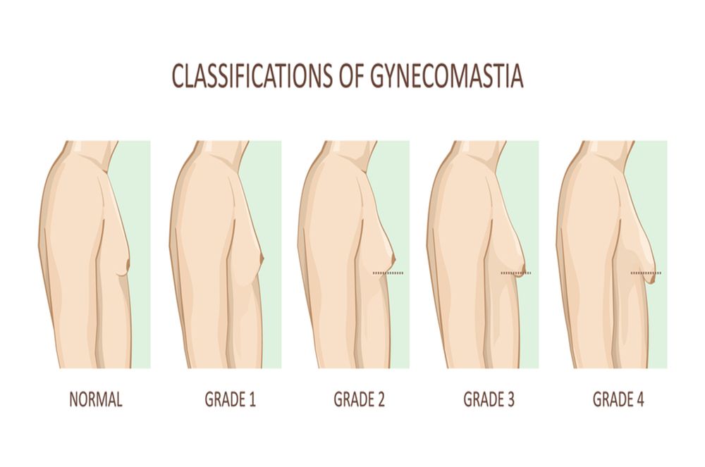
The staging of a progressive condition helps the physician in interpreting the current condition of the patient and also helps in estimating efficiency of treatment plans. Upon severity and degree of enlargement, gynecomastia has been divided into four stages. These stages may also roughly correspond with histological changes.
Stage 1:
There is small breast enlargement without significant skin excess.
The nipple in this stage appear puffy and conical in shape. The excess breast tissue can be distinguished on the basis of color, from the surrounding normal tissue. Diagnosis and surgical treatment at this stage have a higher chance of producing desired results.
Stage 2a:
There is moderate enlargement without skin excess.
Though this stage is also inappreciable under clothing, the excess tissue appears firm and can cause discomfort in the pectoral region. The extent now involves a wider area of the chest.
Stage 2b:
Tissue enlargement is moderate which also comes with some skin excess.
Also called grade 3, this is the stage where sagging of the enlarged breasts is noted due to greater skin laxity. It can be appreciated even under loose clothing.
Stage 3:
This stage is characterized by a marked enlargement in breast size, and excessive skin.
Also known as grade 4, gynecomastia in this stage acquires the mass and drooping similar to the female breasts. The treatment option available at this stage is liposuction along with gland excision.
Another grading system
Another grading system takes into account, the hypertrophy of the breast structure, and also the resultant drooping. The formalized scale is: [8]
- Grade 1: breasts weigh less than 250 g and there is no sagginess.
- Grade 2: breasts weigh between 250-500 grams, with no sagginess.
- Grade 3: breasts weigh more than 500 g with minor ptosis, or drooping.
- Grade 4: breasts have severe hypertrophy with severe ptosis.
Histological stages
Breasts also undergo histological changes, besides changes in physical appearance, as is explained by the grading system above. Though there is no grading system based on histological changes accompanying gynecomastia, these changes are of significant importance in finding the nature of the patient’s condition. Histological pictures are viewed via a mammogram. Mammography uses x-ray technology to visualize the internal changes of the breast tissue.
Histological picture can be either
- Florid: characterized by ductal proliferation of the glandular breast tissue. It gives a nodular appearance in the subareolar tissue when visualized in a mammogram. This histological picture is seen in early phases of gynecomastia for approximately 1 year.
- Fibrous: is characterized by the fibrosis of the tissue rather than ductal proliferation. On a mammogram, it appears as a density in the area surrounding the nipple. It signifies a late stage of gynecomastia. It is also known as quiescent phase and is a characteristic of gynecomastia lasting for more than a year.
Or intermediate: characterized by both fibrous and ductal proliferation. On a mammogram, there are both, a nodular appearance and diffuse fibrosis of surrounding stroma. This histological picture can be a result of taking exogenous estrogen.