How Osteoarthritis Is Diagnosed?
Proper and timely diagnosis of a disease is the first step in preventing progression of the disease to a complicated outcome. Diagnosis of osteoarthritis is made by an orthopedic doctor on various grounds.
The first thing a doctor does to investigate the complaints, is take a complete and thorough history of the complaints. The history comprises questions asked about the complaints. The detailed answers provided by the patient provide the doctor with an insight into all the possible diseases that the patient might be suffering from. Under the following headings we can see how the doctor investigates to form a diagnosis of osteoarthritis.
Clinical
Clinical investigation means investigation of the symptoms of osteoarthritis like pain and stiffness in the joints. The doctor may proceed by asking questions along the following lines:
- The onset, duration, and progression of pain, and whether the pain is aggravated by rest or motion.
- Any complaint of stiffness in joints, especially in the morning.
- If the patient has noticed any restricted mobility, limitation in movement etc. or any increased predicament at work which can be attributed to the symptoms.
- Any other symptoms like body aches, fever, palpitations etc. These help to rule out other causes of joint pain like rheumatoid arthritis, gout, knee injury etc.
- Any previous injury to the joint.
- Any incidence of similar complaints in blood relatives, occupational history etc.,
- Any specific dietary protocol that the patient is on.
The doctor will then examine the joint. Any signs of tenderness or swelling in the joint capsule will be noted. Based on the findings in this phase, further investigations can be ordered.
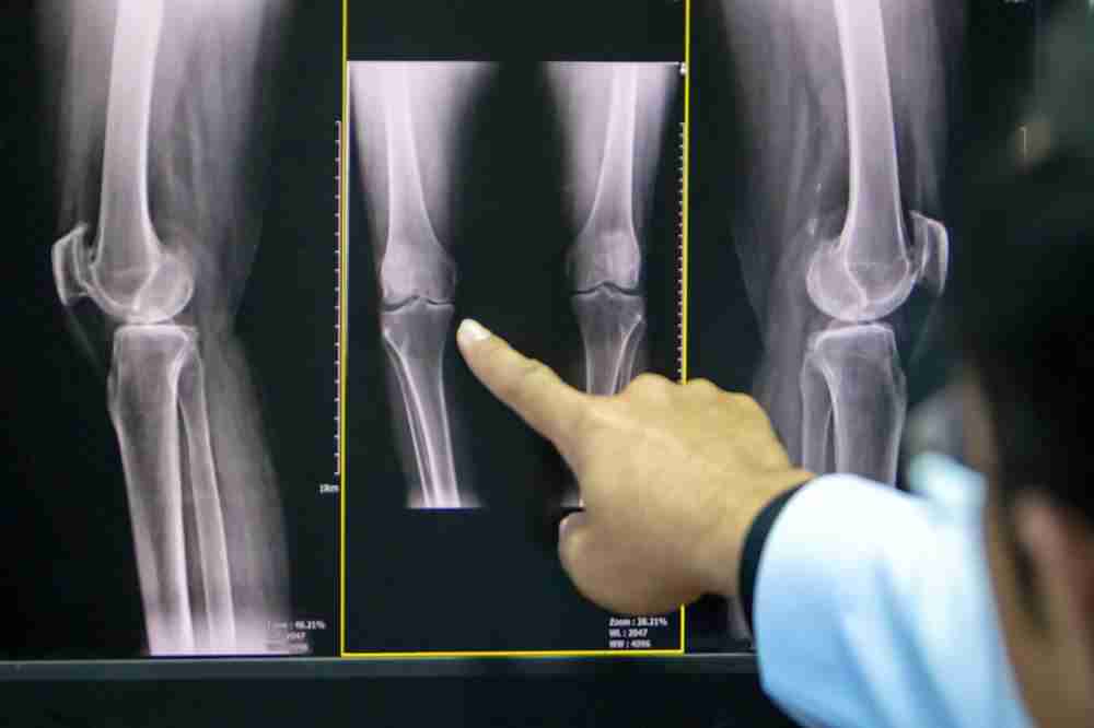
Radiological
The initial investigation in any bone or joint disease is most commonly an x-ray. A plain film radiograph of the joint can show the signs of osteoarthritis. However, the doctor may order x-rays in more than one view to better assess the situation. The following signs are noted in the radiograph:
- The joint space, if it is reduced, is considered a bad sign which can lead to sclerosis of the bone ends.
- The presence of any bony outgrowths or spurs at the joint capsule.
- Any swelling of the joint capsule, which may be a sign of early inflammation.
For accurately monitoring the progression of the disease, multiple radiographs may be ordered from time to time. The radiographic evidence, not always align with clinical symptoms. The patient may report worsening of symptoms, but the radiographs may not indicate a significant and corresponding worsening of the structures.
Classification of osteoarthritis:
On the basis of radiographic imaging, osteoarthritis is classified by Kellgren and Lawrence system:
- Grade 0 means no radiological signs seen
- Grade 1 shows a doubtful picture.
- Grade 2 shows minimal, but distinct signs of bony spur and/or joint space constricting.
- Grade 3 shows a moderate degree osteophyte formation, joint space narrowing and bone deformity
- Grade 4 is characterized by severe bony spurs, joint space sclerosis and bone deformity.
Some blood sampling and sampling of the joint fluid may be done in order to rule out other similar conditions.