Osteoarthritis In all Parts Of The Body
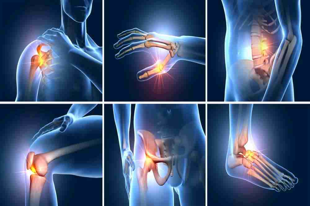
Osteoarthritis can involve multiple joints in the body. The ratio of incidence may differ according to the causative factors like gender. There are also slight differences in radiological findings, and clinical manifestations. We can view them one by one:
Knee
The knee joint is one of the most commonly affected joints due to osteoarthritis. According to a report, 37% of patients over 60 years of age had osteoarthritis of the knee joint. It is also slightly more common in females. Why osteoarthritis attacks, knee joint the most is due to its anatomical location and physiological function. It bears the burden of the entire trunk and upper limbs and its stability is crucial to support proper functioning of the lower limbs.
Abnormal knee alignments like in bow legs called ‘varus’, and knock knees called ‘valgus’ can also lead to increased chances of getting osteoarthritis. Varus alignment increases the chance of osteoarthritis in the medial side of the knee joint. Valgus alignment predisposes to lateral side OA. This is again because of increased force of action passing on the consecutive sides.
Radiographs in anterior-posterior position and lateral views can help in checking for abnormal signs of osteoarthritis. OA can affect either the joint between the thigh bone and leg bones or involve the knee plate, patella. Mostly the joint between the patella and the femur is the first to be affected.
Hip
The hip joint is nearly the second most affected joint from osteoarthritis. A study shows 27% of patients above 45 years to be involved in OA of the hip joint. The hip joint is formed between the head of the large femur bone and its socket in the pelvic bone. The OA of the hip joint may cause pain in the area around the groin and may even radiate to the back and knee due to loss of support at the junction of the trunk and lower body.
The osteoarthritis changes can be visualized on plain x-ray from the front. The upper and outer side of the joint bearing the most pressure and weight is most commonly affected. At this site osteophytes or bony outgrowths can be seen. Bony can also be deformed at this site.
Hands and fingers
Another widely affected site from arthritis is the hand. About 27% of all OA patients have the disease in their hands. Among a number of joints in the hand, the joint where the thumb joins the wrist, and the joints in the fingers are most commonly affected. In the fingers, the distal joints nearer to the nail bed may get affected which give rise to Heberden’s nodes. The joint in the middle of the finger gives rise to Bouchard’s nodes. These nodes are swellings due to bony outgrowths or spurs as a reaction to degrading cartilage in osteoarthritis.
Osteoarthritis of the hands, unlike that of other joints, takes away the liberty of fine motor skills from the patient. The patient will have difficulty in doing chores like buttoning shirts, writing for longer periods, typing or drawing.
As mentioned above, the nodes cause the fingers to become wobbly and stiff. Similarly, if there are bony outgrowths involving the bones that join together to form the wrist, it can complicate into carpal tunnel syndrome. The bony spurs compress the nerves passing near these joints. This can lead to pain like piercing pins in the hands. The symptoms involved with decreased motility at wrist include compromised grip strength, rendering a variable number of tasks impossible to accomplish like holding a brush or handling the utensils etc.
Spine, neck and the back
The joints in the spinal column are most commonly affected in old age, obeying the general trend of incidence of osteoarthritis. The individual bones making the spine are called vertebra. The facet joints between these vertebrae are most commonly affected by osteoarthritis.
The consequences of osteoarthritis in the spine can be more worrisome as this bony column protects the spinal cord, an extension of the brain. The nerves leave the vertebral canal through passages between these bones. If these bones get damaged in osteoarthritis, the bone irregularities and spurs can compress these nerves which supply farther areas in the body. So the symptoms may vary from localized pain in the neck or back or lower spine on twisting, bending or straightening to weakness, numbness, sensory loss in the limbs and other body parts. These extra symptoms will lead to an additional burden of disease on the quality of life.
Radiographic findings can show deterioration of bone blocks, building the spine, decrease in the articular cartilage between these bones causing fusion and bony spurs. MRI scans are also useful in detecting spinal cord compression.
Shoulder
The shoulder joint is not as commonly affected as the above mentioned joints. It is a more likely outcome of sports involving overhead movements, or direct falls or accidents on the shoulder joint. The shoulder joint is made up of the head of the humerus in its socket in the shoulder bone. With progressive osteoarthritis, the bone deformations may lead to loss of normal ball and socket anatomy.
The radiographs can be taken in the axillary view, which accurately demonstrate any changes in the joint space, any swelling of the joint capsule, and may also rule out shoulder dislocation as a likely cause of pain.
Ankle and foot
In the foot, the big toe is by far the most common site of osteoarthritis. It may be caused by hard kicking or something falling on this joint, or twisting and bending of joints at unnatural angles.
The complicated picture of osteoarthritis in the big toe can result in Hallux valgus that is the inward turning of the big toe due to bony outgrowth on the lateral side of the toe. This outgrowth is also called a Bunion. Complete erosion of the cartilage in this joint can result in fixation of the joint called Hallux rigidus.
If we draw a line of balance from the trunk to the ground, the lowest point bearing the pressure is the ankle joint. The tendons at this joint may thin out putting more strain at the joint rendering the patient bedridden, unable to put the feet on the ground. Osteoarthritis in feet can be a likely outcome of shoes like flats and high heels, which put the feet at unnatural angles.