Diagnosis of Osteoporosis

Osteoporosis is a slowly progressive disorder that compromises bone health over time. If there is,
- A non-traumatic fracture
- In an elderly male or post-menopausal women, or a malnourished child
- Pain in the bones
- General changes in the posture and physique
- Changes in the gait and body frame
- A family history of osteoporosis
Laboratory Diagnosis Of Osteoporosis

For confirming a diagnosis, the most common and reliable test available is the bone density test.
Bone density test
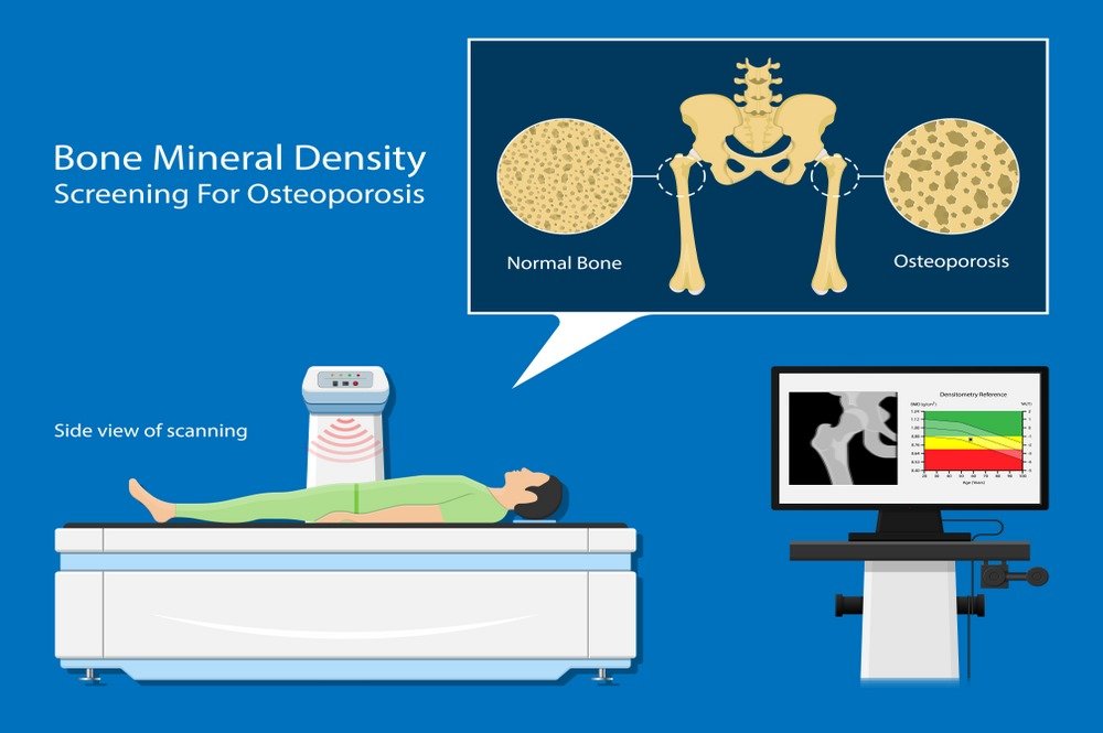
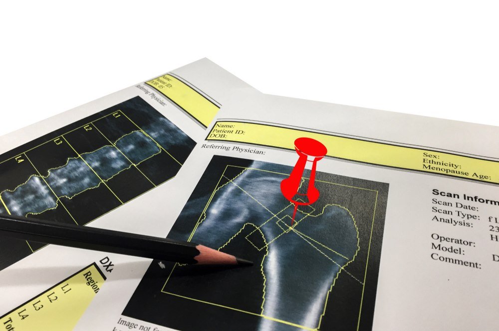
This non-invasive procedure can detect bone fragility before osteoporosis presents itself with a fracture. A central DXA machine uses the principle of dual x-ray absorptiometry to scan the axial skeleton. It uses only mild radiations to develop an image of the bone and detect the degree of bone mineralization. Primarily the scan of the hip or spine is advised, as these are most likely to suffer from an osteoporotic fracture. Regular x-rays can be used to locate the fracture, but they cannot show bone density, until in late stages of osteoporosis.
The interpretation of this test is upon the T-score, which compares the bone density of the patient with that of a normal 30-year-old individual
- A T-score of -1.0 or above indicates normal bone density.
- A T-score value that lies between -1.0 to -2.5, indicates lowered bone density, or a possible case of osteopenia.
- A T-score value Of -2.5 or below is diagnostic of osteoporosis.
By the T-score value, the physician can map out further directions. It can help in affirming whether the patient need to start anti-osteoporosis medication or simple lifestyle change scan improve bone health.
Peripheral Bone Density Test

In case, the central DXA is not available, or cannot be done in patients weighing more than 300 pounds, the peripheral bones in the appendicular skeleton; the wrist, fingers, heel, or arm can be evaluated. The following tests are used
- Peripheral dual x-ray absorptiometry (pDXA)
- Quantitative ultrasound (QUS)
- Peripheral quantitative computed tomography (pQCT)
These are screening tests and not diagnostic tests for osteoporosis. They only give an estimate of the bone density and mineralization and help in assessing the bone status of the patient. These tests give a clear idea as to the requirement of further tests.
FRAX Score

A FRAX score sheet is a developed questionnaire that predicts the risk of fracture in the next 10 years. It is also available as a free online service. It takes into account the following factors which help evaluating the bone density:
- Age
- Sex
- Previous history of bone fractures
- Family history
- Height
- Weight
- Glucocorticoid
- Smoking history
- Alcohol use
- Secondary osteoporosis
FRAX score is given in percentage, and is interpreted as percentage risk, or 10-year probability of two variables, the major osteoporotic fracture, and the hip fracture.
Blood and Urine Tests

Blood and urine tests can be requested by the doctor to identify any underlying condition that may cause bone loss. This includes screening for calcium levels. Samples are also tested for the concentration of sex hormones, thyroid and parathyroid hormones.

