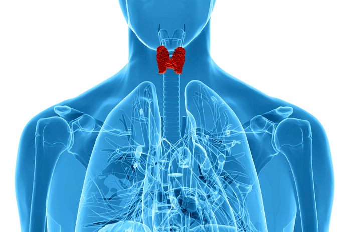
Diagnosis Of Thyroid Cancer
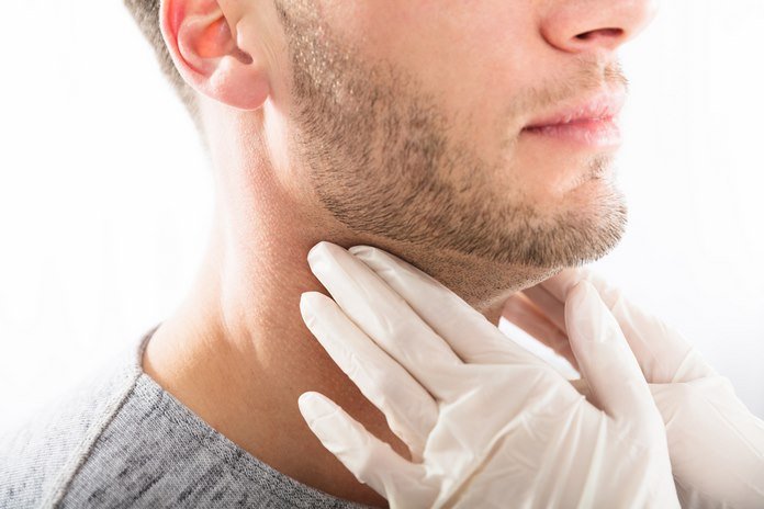
Physical examination and medical history of the patient:
The doctor will perform a physical examination to check for any signs and symptoms of thyroid cancer. Doctors examine the areas like the neck, thyroid gland, lymph nodes in the neck, and throat for swelling and abnormal growth. The doctors ask the patients questions about symptoms and risk factors, along with medical history. Doctors also ask about family history of thyroid cancer. The patients having family history are at more risk of developing thyroid cancer.
Blood Tests
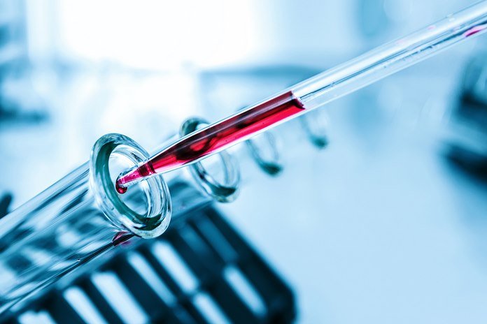 Tumor cells secrete some substances that accumulate in the blood; these substances are known as tumor marker cells. The presence of these substances in the blood indicates the presence of cancer in the body. Doctors use blood tests to detect the presence of these substances in the blood and to monitor the patient’s treatment. The blood tests also check the performance of the thyroid gland by measuring the level of hormones in the blood produced by the thyroid gland.
Thyroid Hormone Test
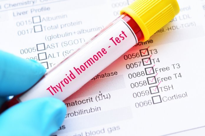 The thyroid gland produces triiodothyronine (T3) and thyroxine (T4). These hormones regulate many functions in the body. Blood tests measure the levels of these hormones in the blood. Higher or below normal levels of these hormones in the blood indicates an abnormality of thyroid gland functioning.
TSH Levels
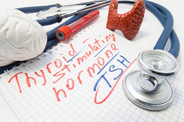
The pituitary gland produces thyroid-stimulating hormone (TSH) in response to the need for thyroid hormones. This test gives the level of thyroid-stimulating hormone in the blood.
Thyroglobulin Levels
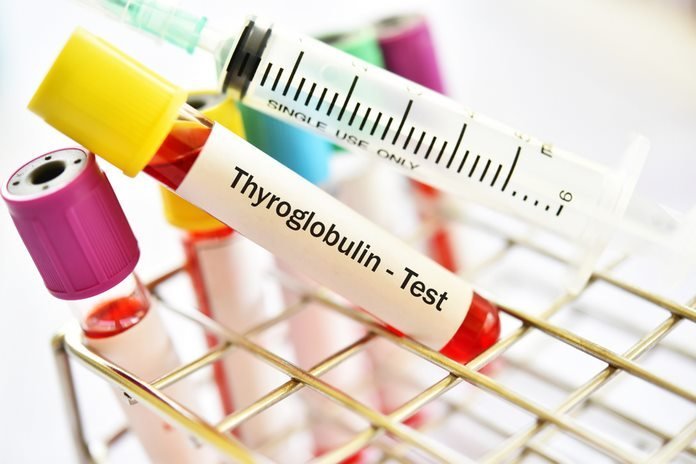 thyroid cells, as well as cancerous thyroid cells, produce thyroglobulin. The high levels of thyroglobulin in the blood after treatment indicate the presence of remaining thyroid cells, which may be cancerous. This test usually performs after treatment to check the presence of thyroid cells that escapes the treatment.
Calcitonin Levels
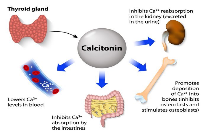
C cells of thyroid produce a hormone known as calcitonin that helps to regulate the use of calcium in the body. The medullary thyroid cancer develops in the C cells of the thyroid. Levels of calcitonin in the blood help to diagnose medullary thyroid cancer. Blood calcium levels and carcinoembryonic antigen levels checked as well.
Ultrasound
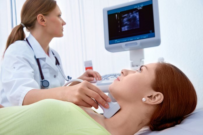 This test uses high energy sound waves to create images of the thyroid gland. The ultrasound takes less time, painless, and does not expose the body to radiations. Normally ultrasound takes 15 to 20 minutes and makes the picture on a computer screen. Ultrasound can detect if the nodule present in the neck is solid or a cyst filled with fluid. This method uses a gel to spread on the area need to examine, and a probe moved on the skin. This method is the best way to get detailed information about the internal organs.
Biopsy
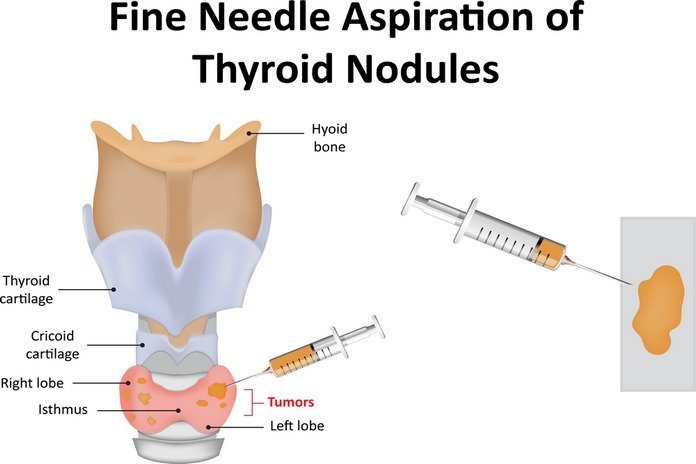
Doctors use this method to make an exact diagnosis of cancer. In this procedure, a small amount of tissue removed by needle examined under the microscope to check for the presence of cancerous cells. There are two methods to perform a biopsy for the diagnosis of thyroid cancer.
Fine needle aspiration (FNA): Doctors use FNA to check if the nodule is cancerous or not. This procedure uses a small thin needle that is inserted into the skin to collect cells and fluid. Fine needle aspiration may be done at different parts of the body. FNA is normally performed in a clinic or hospital. The patient may be given a small amount of local anesthetic before the biopsy. A positive or negative test determines the presence or absence of cancerous cells in the collected sample.
Surgical Biopsy
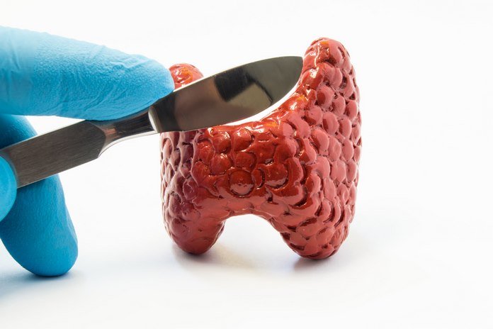 doctors use surgical removal of the nodule or lobe of the thyroid gland if the results of fine-needle aspiration are not clear—the surgical biopsy normally performed at the hospital under local anesthesia. After the biopsy, patients need to remain in the hospital for 1 to 2 weeks.
Radioiodine Scan
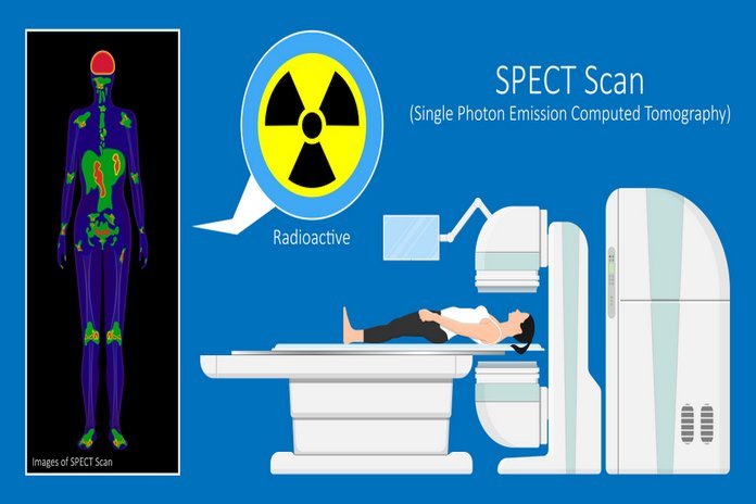 In this procedure, the patient is given or injected with a small amount of radioactive iodine I-131 or I-123. Thyroid cells absorb the iodine, often known as a tracer. This test checks if the nodule or lump in the neck is cancerous or not. The absorption of iodine in the cells helps to appear them on the scan and allows the doctor to check for abnormality in the cells as compared to normal cells.
X-Ray
 X-ray technique creates images of the internal body structure by using radiations. Usually, doctors use the x-ray to check if cancer has spread to other body parts such as chest and lungs.
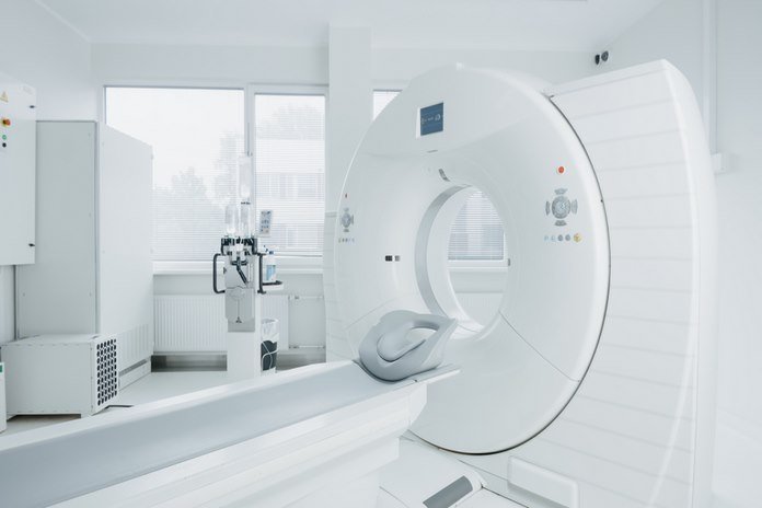
CT Scan
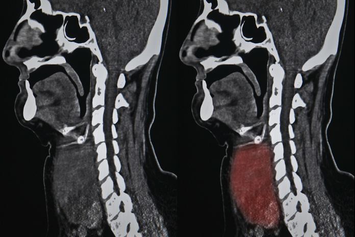 This technique creates three-dimensional images of the body on the computer by using an x-ray. The patient may be given a small amount of contrast material before scan for improved detailing of the images produced by the scan. Doctors use a CT scan to examine parts of the body that cannot be examined by ultrasound.
PET Scan
 This technique uses a small amount of sugar injected into the body of the patient. The cells with high energy demand absorb radioactive sugar. The cancer cells absorb more sugar than normal cells because of their high demand for energy. The scanner produces images of the body by detecting the sugar absorbed by the cells. This technique usually combined with a CT scan and known as a PET-CT scan.